Melanoma is the most lethal form of skin cancer, arising from melanocytes. This guide delves into its causes, types, early detection using the ABCDE rule, staging systems like Breslow Depth and AJCC, and modern treatment options including surgery, immunotherapy, and targeted therapy.
Introduction
This article delves into the most lethal form of skin cancer. Melanoma is skin cancer arising from melanocytes, it is responsible for the majority of skin cancer-related deaths. Its aggressive nature and potential to metastasize demands early recognition and aggressive intervention. Prolonged and excessive exposure to UV radiation from the sun, once again puts people at high risk for developing melanom.
Etiology of Melanoma
Melanoma primarily arises from the cumulative effects of exposure to ultraviolet (UV) radiation from the sun or tanning beds, which damages the DNA within skin cells. However, the disease's complexity extends beyond UV exposure, with other contributing factors such as having fair skin, a history of severe sunburns -especially during childhood and adolescence - a family history of melanoma, the presence of numerous moles, a weakened immune system, and hormonal changes like pregnancy. (1)
Types of melanoma
-
Superficial Spreading Melanoma (SSM): This is the most common type of melanoma, accounting for approximately 70% of cases. It typically starts as a flat or slightly raised lesion with irregular borders. Over time, it may grow horizontally before penetrating deeper into the skin.
 Figure 1.
 Figure 2.
 -
Nodular Melanoma (NM): This is a particularly aggressive variant, representing approximately 15-30% of cases. It typically presents as a dome-shaped nodule, often dark in color, and is prone to bleeding and ulceration. Unlike other forms, NM often lacks the typical signs of asymmetry, irregular borders, and color variation. It tends to grow quickly and it is frequently diagnosed at a later stage, contributing to a less favorable prognosis.
 Figure 3.
 -
Lentigo Maligna Melanoma (LMM): This type of melanoma usually affects older individuals with a history of extensive sun exposure. LMM typically develops slowly and initially appears as a flat, tan or brown patch on the skin. Over time, it may become darker or develop irregular borders. It has a better prognosis compared to other subtypes when detected and treated early.
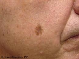 Figure 4.
 -
Acral Lentiginous Melanoma (ALM): This subtype primarily affects the palms of the hands, soles of the feet, or under the nails. It is more common in people with darker skin tones and is not strongly associated with UV exposure. It often starts as a dark spot or discoloration and may be mistaken for other non-cancerous conditions. ALM is typically diagnosed at a more advanced stage due to its location and diagnostic challenges, contributing to a less favorable prognosis. Interesting fact: Bob Marley died from an ALM at age 36.
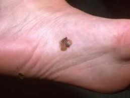 Figure 5.
 -
Amelanotic Melanoma: Amelanotic melanoma is a subtype of melanoma that lacks the typical dark coloration due to a lack of pigment production. It can appear as a flesh-colored or pinkish bump or lesion, making it easily mistaken for other benign skin conditions. Due to the difficulty in diagnosis, amelanotic melanoma is often detected at later stages, impacting prognosis.
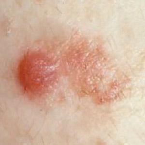 Figure 6.
 -
Mucosal Melanoma: These can occur on any mucous membrane, such as those lining the mouth, nasal passages, gastrointestinal tract, and genitalia and can appear as pigmented or non-pigmented lesions. They tend to be diagnosed at later stages due to their hidden location and diverse appearance and thus have a poor prognosis.
-
Ocular melanoma: Ocular melanoma is the second most common type of melanoma after cutaneous and is the most common primary intraocular malignant tumor in adults.
-
Desmoplastic Melanoma: This is a rare subtype that tends to occur on sun-exposed areas, such as the head and neck. It is characterized by a fibrous or scar-like appearance and can be challenging to diagnose. Desmoplastic melanoma has a higher tendency to spread along nerve pathways. (2)
Early Detection and Diagnosis
Melanoma can appear as a new mole or develop within an existing mole. It is crucial to recognize the warning signs and seek medical attention if any lesion exhibits the "ABCDE" characteristics: asymmetrical shape, irregular borders, color variation, diameter larger than 6 millimeters, and evolving characteristics. Other symptoms may include itching, bleeding, or a sore that does not heal. Not all melanomas exhibit these symptoms, so any suspicious changes in the skin should be promptly examined by a healthcare professional.
As mentioned, in the pursuit of better melanoma outcomes, early detection is vital. Dermoscopy, a non-invasive technique, enhances diagnostic accuracy by magnifying skin lesions and recording images for follow-up comparison to detect subtle changes.
 Figure 7. Image demonstrating a comparison of lesion on Molemax system (3)
Any suspicious lesion should be biopsied or completely excised whenever possible.
How Deep Is It? Understanding Melanoma Staging
When doctors diagnose melanoma (a serious type of skin cancer), one of the most important things they look at is how deep the cancer has grown into the skin. This helps predict how serious it is and what kind of treatment may be needed.
Breslow Depth: How Far Has It Gone?
Think of the Breslow Depth like a ruler that measures how deep the melanoma cells have spread into your skin—from the very top layer (epidermis) down into the deeper layers.
- The deeper the melanoma goes, the more serious it can be.
- This depth helps doctors decide on the next steps in treatment.
Although the Breslow Depth is still used, today most doctors use a more advanced system called the AJCC staging system (more on that below).
Clark Level: An Older System
Before Breslow Depth and AJCC staging became common, doctors used something called the Clark Level. It described which layers of the skin the melanoma had reached:
Level 1 Cancer is only in the very top layer of skin (epidermis).
Level 2 It's moved into the top part of the second layer (dermis).
Level 3 It's going deeper into the dermis.
Level 4 It's reached the lower part of the dermis.
Level 5 It has spread into the fat beneath the skin.
Over time, doctors found that Clark Levels weren't always reliable, so they've mostly been phased out.
Today's Standard: AJCC Staging
Now, the most widely used method for staging melanoma is the AJCC system (developed by the American Joint Committee on Cancer). It uses the TNM system:
- T = Tumor (How thick is it? Is it ulcerated?)
- N = Node (Has it spread to nearby lymph nodes?)
- M = Metastasis (Has it spread to other parts of the body?)
This system helps doctors determine how advanced the cancer is and what treatment options will be most effective. It also helps compare outcomes between patients at the same stage.
Treatment: How Is Melanoma Treated?
The way melanoma is treated depends on how early it's caught, where it's located, how big it is, and your overall health. But one thing is clear: the earlier it's found, the better the outcome.
Surgery: The First Step
In most cases, surgery is the main treatment. If a suspicious spot turns out to be melanoma, doctors usually remove it along with some of the healthy skin around it. This helps make sure all the cancer cells are gone. (4)
The amount of skin that needs to be removed depends on how deep the melanoma goes:
- If it's very thin, a small margin is enough.
- If it's deeper, doctors will remove a wider area around it.
Checking the Lymph Nodes
If the melanoma is 1 millimeter deep or more, doctors often check to see if it has spread to nearby lymph nodes. This is done with a special test called a sentinel lymph node biopsy. If cancer cells are found there, more tests and possibly more treatment will follow. (5)
What if the Melanoma Has Spread?
If melanoma has spread beyond the skin (called metastatic melanoma), treatment becomes more complex. Fortunately, there have been big breakthroughs in recent years. There are two main kinds of newer treatments:
- Immunotherapy: These medicines help your body's own immune system recognize and attack the cancer. Some people see their tumors shrink or disappear, sometimes for years. However, these treatments can cause serious side effects, so they're closely monitored.
- Targeted therapy: About half of all melanomas have a change in a gene called BRAF. If your melanoma has this gene change, doctors may give you special drugs that "target” it. These medications are often used in combination to be more effective.
In some cases, doctors might combine immunotherapy with targeted therapy, but choosing the right treatment depends on many factors and should always be handled by a specialist. (6)
What's the Outlook (Prognosis)?
- If melanoma is found early, it's usually very treatable, and many people go on to live long, healthy lives.
- But if it's found later, especially after it has spread, it becomes more difficult to treat and the chances of cure decrease.
This is why checking your skin regularly and catching changes early is so important.
What Happens After Melanoma Treatment?
Even after treatment, follow-up care is crucial—because once you've had melanoma, you're at a higher risk of developing it again than someone who never has. This doesn't mean you'll definitely get it back, but it does mean you need to stay alert and regularly check in with your doctor.
Here's what follow-up care usually looks like, depending on the stage of your melanoma:
Stage 0 (also called "in situ")
- This is the earliest and least serious stage of melanoma.
- You'll need a full skin check once a year for life.
- Do monthly self-checks at home to spot anything new or changing.
- You won't need regular blood tests or scans unless something unusual comes up.
Stage I to IIA
- These are still early-stage melanomas, but they've gone a bit deeper into the skin.
- Doctors will check your skin and general health every 6 to 12 months for the first 5 years, and then once a year after that.
- Keep doing your monthly self-exams at home.
- Routine blood work or scans aren't needed unless you're having symptoms.
Stage IIB to IV (no evidence of disease)
- These stages involve deeper or more advanced melanoma, but in these cases, the cancer has been treated and is no longer visible.
- You'll have checkups every 3 to 6 months for the first 2 years, then every 3 to 12 months for the next 2 years, and once a year after that.
- Imaging scans (like CTs or MRIs) may be done, but only if needed based on your symptoms or doctor's judgment.
- Regular blood tests and scans aren't usually part of long-term follow-up unless you're in a clinical trial. (7)
Don't Forget Your Own Monthly Skin Checks
You know your body best. Make it a habit to look over your skin every month—head to toe—and talk to your doctor if you see anything new, changing, or just doesn't look right. Early detection can save lives.
Conclusion
Melanoma is one of the most dangerous forms of skin cancer, but it is also one of the most preventable and treatable, especially when caught early. Understanding the risks, recognizing the warning signs, and taking proactive steps like using sunscreen, avoiding excessive sun exposure, and performing regular skin checks can significantly reduce the chance of serious outcomes.
If diagnosed, staying informed, following recommended treatment plans, and attending follow-up appointments are key to improving long-term prognosis. With awareness and early action, melanoma doesn't have to be a life-threatening condition. Protect your skin, stay vigilant, and prioritize your health because early prevention can truly save lives.